Explore web search results related to this domain and discover relevant information.
The skin is the body's largest organ. It serves as a protective shield against heat, light, injury, and infection.
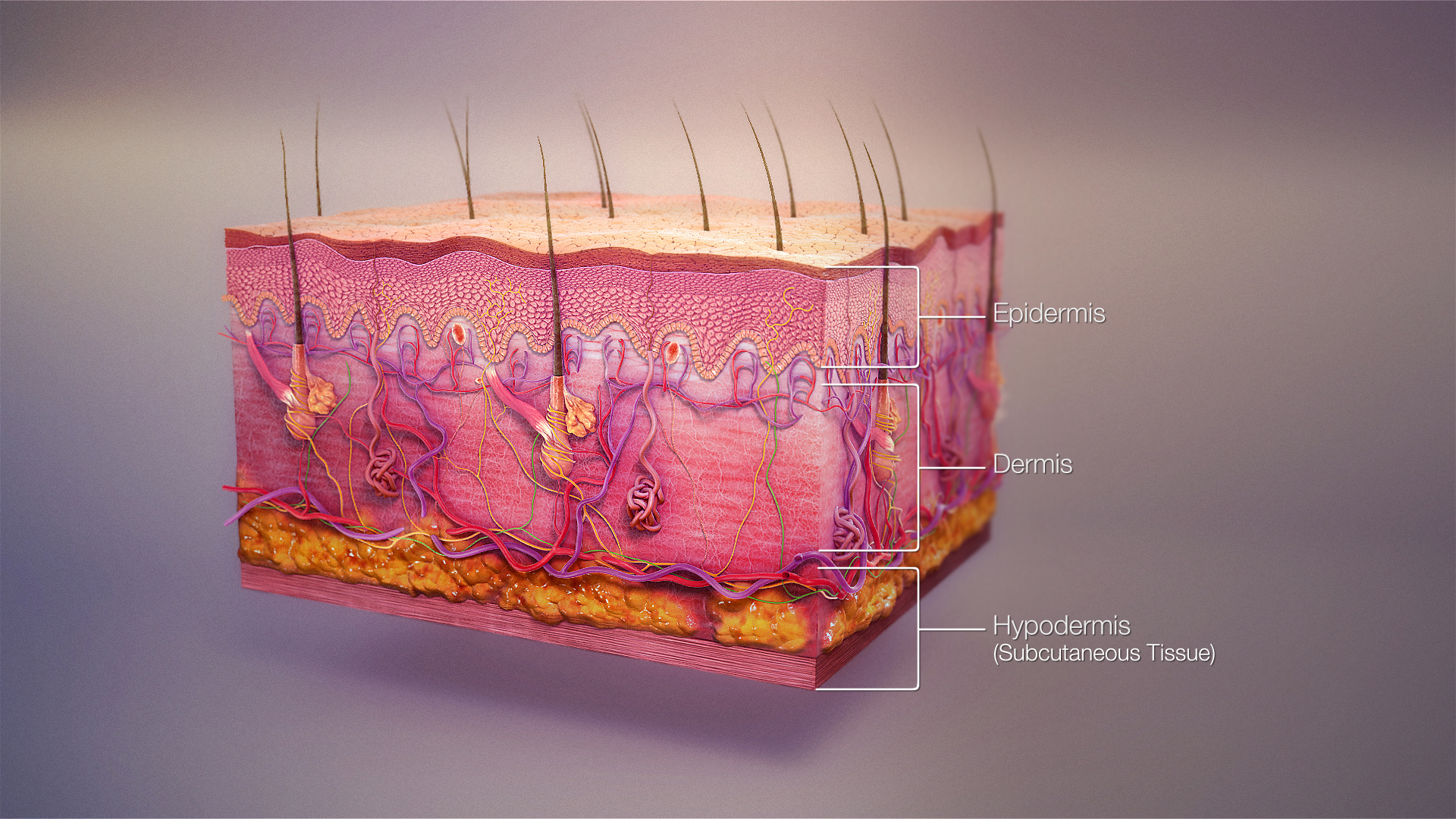
The skin is the body's largest organ. It covers the entire body. It serves as a protective shield against heat, light, injury, and infection.Your skin takes on different thickness, color, and texture all over your body. For example, your head contains more hair follicles than anywhere else. But the soles of your feet have none.In addition, the soles of your feet and the palms of your hands are much thicker than skin on other areas of your body.The skin is made up of 3 layers.

The stratum corneum is composed of keratin and dead keratinocytes (anucleate squamous cells) that form horny scales. This layer has the most variable thickness, especially in callused skin. Dead keratinocytes release defensins within this layer, which are part of our first line of immune defense ...
The stratum corneum is composed of keratin and dead keratinocytes (anucleate squamous cells) that form horny scales. This layer has the most variable thickness, especially in callused skin. Dead keratinocytes release defensins within this layer, which are part of our first line of immune defense mechanisms.[6][7]Langerhans cells are dendritic cells that act as the skin's first-line cellular immune defenders and are crucial for antigen presentation. Special stains allow visualization of these cells in the stratum spinosum. Langerhans cells are of mesenchymal origin, derived from CD34-positive bone marrow stem cells, and are part of the mononuclear phagocytic system.The skin-associated lymphoid tissue is a significant component of the immune system, aiding in preventing infections, as even minor skin breaks can lead to infection. Langerhans cells are part of the adaptive immune system, presenting foreign antigens encountered in the skin to T cells.Regulation of homeostasis: The skin plays a vital role in maintaining body temperature and water balance. This organ regulates heat exchange with the environment, particularly through the blood vessels and sweat glands.
The blood supply originates from ... tissue and dermis, respectively.[4] An extensive lymphatic framework runs alongside many of the skin’s blood vessels, particularly those attached to the venous end of the capillary networks.[3] See Image....
The blood supply originates from an extensive network of larger blood vessels and capillaries that extend from regional branches of the systemic circulation to local sites throughout subcutaneous tissue and dermis, respectively.[4] An extensive lymphatic framework runs alongside many of the skin’s blood vessels, particularly those attached to the venous end of the capillary networks.[3] See Image.Dermatomes are areas of skin mainly supplied by a single spinal nerve. Eight cervical nerves contribute to the dermatomes (except for C1), 12 thoracic nerves, five lumbar nerves, and five sacral nerves. Each of these nerves relays sensation (including pain) from a particular region of the skin to the brain.[4]Skin infections include cellulitis, erysipelas, and impetigo caused by staphylococcal and streptococcal bacteria.[3][4] ... In the human body, the sensory fibers carrying pain stimuli are arranged into dermatomes, which are segmentally distributed across the entire body surface (illustration below). Dermatomes develop embryologically. A dermatome functionally represents how sensory information (e.g., pain) travels from a particular skin receptor type (e.g., nociceptor) to the corresponding peripheral nerve then connects to a specific spinal nerve (cervical; C1–C8, thoracic: T1–T12, lumbar: L1–L5 and sacral: S1–S5), reaching the spinal cord where the signals ultimately ascend to the brain.[6][7][8]Regulation of Temperature. Skin participates in thermal regulation by conserving or releasing heat and helps maintain the body’s water and homeostatic balance.[1][2]
The epidermis is the most superficial layer of the skin and provides the first barrier of protection from the invasion of substances into the body · The epidermis is subdivided into five layers or strata:
Pediatric Core Concepts Dermatology Chapter

The integumentary system has five components: skin, hypodermis, hair, nails, and exocrine glands. The different components work together to maintain health and well-being for the rest of the body. ... Protection. The epidermis and dermis form a physical barrier that protects all the internal body parts...
It offers protection for the skin, increases sensory function, and helps regulate body temperature. Nails: Nails are layers of keratin that form hard plates. They grow over the tips of the fingers and toes. Nails protect the digits and aid in dexterity. Glands: Four types of glands make up integumentary system parts: sudoriferous, sebaceous, ceruminous, and mammary glands.Find out what you need to know about the integumentary system and discover how it may affect your health.The integumentary system is the physical system that forms the barrier between the external environment and the internal systems of the body. In humans, this system consists of skin, hair, nails, and related glands.The skin protects against negative effects of your environment such as heat, cold, and sun exposure, and it prevents many foreign substances, such as bacteria, viruses, and environmental contaminants, from getting inside the body. The hypodermis layers beneath the skin provide additional cushioning for internal organs.

Protecting and repairing the most visible part of the body. ... This Nature Outlook is editorially independent, produced with financial support from LEO Pharma. About this content. The word skin has come to stand for superficiality and lack of depth, but human skin defies such connotations.
For one thing, it is enormous, accounting for roughly 15% of a person’s body weight. Skin is also remarkable for its diverse roles: protection against pathogens, temperature regulation and, of course, sensation. Like any part of the body, skin is fallible to disease.Skin interacts with other parts of the body in complex and sometimes mystifying ways. People affected by atopic dermatitis, for example, are at greater risk of a range of mental-health disorders.How research is protecting and repairing the most visible part of the body.Gene therapies, for example, are yielding remarkable results in correcting a rare disorder that results in fragile ‘butterfly skin’, enabling children with the skin condition to live active lives. People with psoriasis, who have long been faced with the need to take antibody medications their whole lives, could soon find longer-lasting relief in more-durable treatments.

In Java Edition, any pixels on the bottom layer of uploaded skins will become fully visible if not already, but their red, green, and blue values will be rounded to parts per opacity value instead of parts per 255 (then will be rounded back into parts per 255), or will appear black if opacity is 0.
For example, if a pixel's red, green, and blue values are 255, 13, 142, with an opacity of 2, their red, green, and blue values will be rounded to parts per two, as one of 0/2, ~1/2, or 2/2 (0, 128, or 255), and become 255, 0, 128, with an opacity of 255. Pixel size of the 2nd layer on body, arms and legs is 0.25 pixel bigger than the skin pixel (inner layer).The Marketplace, which officially hosts a large number of skin packs by Mojang Studios and their partners.The modern layered skin template; every body part can have a second layer, marked by the darker palette areas.Skins refer to the textures that are placed onto the player character model. Skins can be easily created using a skin editor.
The human skin is the outer covering of the body and is the largest organ of the integumentary system. The skin has up to seven layers of ectodermal tissue guarding muscles, bones, ligaments and internal organs. Human skin is similar to most of the other mammals' skin, and it is very similar ...
In terms of surface area, the skin is the second largest organ in the human body (the inside of the small intestine is 15 to 20 times larger). For the average adult human, the skin has a surface area of 1.5–2.0 square metres (15–20 sq ft). The thickness of the skin varies considerably over all parts of the body, and between men and women, and young and old.The subcutaneous tissue (also hypodermis and subcutis) is not part of the skin, but lies below the dermis of the cutis. Its purpose is to attach the skin to underlying bone and muscle as well as supplying it with blood vessels and nerves. It consists of loose connective tissue, adipose tissue and elastin.Aesthetics and communication: others see skin and can assess mood, physical state and attractiveness. Storage and synthesis: acts as a storage centre for lipids and water, as well as a means of synthesis of vitamin D by action of UV on certain parts of the skin.Oily surfaces, such as the face, may contain over 78 million bacteria per square centimetre (500 million per square inch). Despite these vast quantities, all of the bacteria found on the skin's surface would fit into a volume the size of a pea. In general, the microorganisms keep one another in check and are part of a healthy skin.The human skin is the outer covering of the body and is the largest organ of the integumentary system. The skin has up to seven layers of ectodermal tissue guarding muscles, bones, ligaments and internal organs. Human skin is similar to most of the other mammals' skin, and it is very similar ...
Health indicator. From looking at your skin's color, firmness, or texture, you or your doctor can gather clues about your overall health. There are medical terms for various parts of your skin.
The epidermis is the thinnest layer of your skin, but it's responsible for protecting you from the harsh environment. The epidermis has five layers of its own. It also hosts different types of cells. Keratinocytes produce the protein known as keratin, the main part of the epidermis.Protect your skin. Too much exposure to UV radiation (from sunlight) is bad for your skin. Experts note that the parts of your body regularly exposed to sunlight (face, arms, neck) tend to show more visible signs of age than other body parts.Wear sunscreen and sunglasses regularly and cover up those body parts that often get exposed to the sun. When you hear the word elastin, think elastic. This protein is found with collagen in the dermis and gives structure and support to your skin and organs. As with collagen, elastin is affected by aging and environmental factors.You can lose hair from any part of the body, but it's mostly from the face (such as eyelashes) and the scalp. Some immune diseases, as well as having a family member with alopecia, may increase your chance of getting this condition. Most people with alopecia don't have any other health problems beside hair loss. ... Your immune system is overactive, causing skin cells to go into overdrive.
These keratinocytes have the ability ... proteins and lipids, therefore playing a vital role in skin function (Bouwstra et al., 2003). The epidermis can be subdivided into stratum corneum, stratum lucidum (only in some parts), stratum granulosum, stratum spinosum and stratum ...
These keratinocytes have the ability to differentiate and undergo structural and compositional changes, leading to the synthesis and expression of a variety of structural proteins and lipids, therefore playing a vital role in skin function (Bouwstra et al., 2003). The epidermis can be subdivided into stratum corneum, stratum lucidum (only in some parts), stratum granulosum, stratum spinosum and stratum germinativum.Skin injuries are breaks in the skin tissue caused by surgical procedures, genetic irregularities and physical and chemical traumas. These wounds can also be divided into the following categories based on the depth of damage; epidermal, superficial partial-thickness, deep partial-thickness and full-thickness skin wounds (Papini, 2004).However, in deep partial-thickness and full-thickness skin wounds, self-healing is not possible since the skin's epithelial regenerative elements are completely destroyed (Blanpain et al., 2004; Tumbar, 2006).Scaffolds are the backbones of any tissue-engineered skin substitute. They provide a platform for cells during the healing process. The structure, morphology, surface topography and mechanical elasticity of scaffolds play a crucial role in cell metabolic activities (e.g., cell-adhesion, -proliferation, -growth, and -differentiation) for successful neovascularization and complete wound repair. Traditional methods such as solvent casting/particulate leaching, freeze-drying (lyophilization), gas foaming, electrospinning, micro-patterning and micro-molding have been widely used for the fabrication of bioengineered tissue substitutes (Ma et al., 2003; Savoji et al., 2014a, 2016; Thadavirul et al., 2014; Limongi et al., 2015; Monteiro et al., 2015; Poursamar et al., 2015; Hadjizadeh et al., 2016; Ng et al., 2016; Mahmoudi et al., 2017).
The skin’s main layers include the epidermis, dermis and hypodermis and is prone to many problems, including skin cancer, acne, wrinkles and rashes.
Your skin, along with your hair, nails, oil glands and sweat glands, is part of the integumentary (in-TEG-you-MESkin is the largest organ in the body, protecting it from external elements. Skin consists of many layers, made of water, protein, fats and minerals.As the body’s largest organ, skin protects against germs, regulates body temperature and enables touch (tactile) sensations.The skin’s main layers include the epidermis, dermis and hypodermis and is prone to many problems, including skin cancer, acne, wrinkles and rashes.
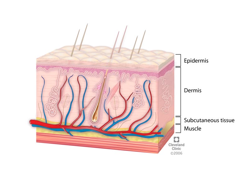

The hypodermis (also called the subcutaneous layer or superficial fascia) is a layer directly below the dermis and serves to connect the skin to the underlying fascia (fibrous tissue) surrounding the muscles. It is not strictly a part of the skin, although the border between the hypodermis ...
The hypodermis (also called the subcutaneous layer or superficial fascia) is a layer directly below the dermis and serves to connect the skin to the underlying fascia (fibrous tissue) surrounding the muscles. It is not strictly a part of the skin, although the border between the hypodermis and dermis can be difficult to distinguish.The first thing a clinician sees is the skin, and so the examination of the skin should be part of any thorough physical examination. Most skin disorders are relatively benign, but a few, including melanomas, can be fatal if untreated. A couple of the more noticeable disorders, albinism and vitiligo, affect the appearance of the skin and its accessory organs.Albinism is a genetic disorder that affects (completely or partially) the coloring of skin, hair, and eyes. The defect is primarily due to the inability of melanocytes to produce melanin. Individuals with albinism tend to appear white or very pale due to the lack of melanin in their skin and hair.The previous edition of this textbook is available at: Anatomy & Physiology. Please see the content mapping table (crosswalk) across the editions. This publication is adapted from Anatomy & Physiology by OpenStax, licensed under CC BY. Icons by DinosoftLabs from Noun Project are licensed under CC BY.

Your skin has three layers that house your sweat and oil glands, hair follicles, melanocytes, and blood vessels · Mayo Clinic does not endorse companies or products. Advertising revenue supports our not-for-profit mission
Learn more about services at Mayo Clinic.

Nerve endings are contained in ... like parts of a puzzle. These projections are especially prominent on the palms of the hands and soles of the feet, where the epidermis is ridged and furrowed in patterns of tiny whorls and loops. These patterns are what form each person’s unique set of fingerprints and footprints. Subcutaneous fatty tissue is the deepest layer of the skin...
Nerve endings are contained in the papillae, tiny projections that fit the dermis to the epidermis like parts of a puzzle. These projections are especially prominent on the palms of the hands and soles of the feet, where the epidermis is ridged and furrowed in patterns of tiny whorls and loops. These patterns are what form each person’s unique set of fingerprints and footprints. Subcutaneous fatty tissue is the deepest layer of the skin.Two sets of glands discharge secretions through the skin. Sebaceous, or oil, glands arise from the walls of hair follicles and produce an oil called sebum that lubricates the skin and hair. Sweat glands, embedded in the subcutaneous layer, are scattered over the body, particularly in the palms and soles.The human body’s largest organ is the skin, or integument. All vertebrates (animals with backbones) have skin, though the covering in each species has different features,…The human body’s largest organ is the skin, or integument. All vertebrates (animals with backbones) have skin, though the covering in each species has different features, such as scales, feathers, or fur (see feather; hair; leather).
Skin Facts · Preventing Sun Damage · Skin Cancer Detection · Sun Safety · Newsletter Archive · JAOCD Archive · Get to Know Your AOCD Historical Series · In The News · Osteopathic Medicine · Corporate Support · Corporate Sponsors · Information for Corporate Sponsors ·
This condition causes a thick, scaly, or crusty skin patch. ... It often appears on parts of the body that receive a lot of sun exposure, such as the hands, arms, face, scalp, and neck.
It may be less visible on darker skin or appear lighter or darker than surrounding tissue. It also causes warm, itchy wheals at the site of contact, which may take on a dry, crusted appearance with repeated exposure to latex. Airborne latex particles may cause cough, runny nose, sneezing, and itchy, watery eyes.There are many skin disorders. Some are temporary, but others are permanent and more serious. Learn about identification, treatment, and prevention.Skin disorders, such as acne and eczema, vary greatly in symptoms and severity. They can be temporary or permanent and may be painless or painful.Some skin disorders have situational causes, while others may be genetic.

Humans shed around 500 million skin cells each day. In fact, the outermost parts of the epidermis consist of
The deepest layer of the skin is the subcutaneous tissue, the hypodermis, or the subcutis. It is not technically part of the skin, but it helps attach the skin to the bones and muscles.Protecting against pathogens. Langerhans cells in the skin are part of the immune system.Recommended skin care for older adults places particular emphasis on moisturizing the skin and keeping it protected from the sun.The skin is our largest organ. Here, we explain what it's made of, what it does, and how it does it. We also cover some common skin conditions.
Epidermal appendages are intradermal ... reepithelialization should the overlying epidermis be removed or destroyed in situations such as partial thickness burns, abrasions, or split-thickness skin graft harvesting....
Epidermal appendages are intradermal epithelial structures lined with epithelial cells with the potential for division and differentiation. These are important as a source of epithelial cells, which accomplish reepithelialization should the overlying epidermis be removed or destroyed in situations such as partial thickness burns, abrasions, or split-thickness skin graft harvesting.Clinically, this extensive horizontal network of vessels allows for random skin flap survival. [34, 35, 36] Studies have further detailed the organization of dermal arteries into discrete arterial units, challenging the traditional concept of a continuous superficial dermal arterial plexus. Research on the cutaneous angiosome of the descending genicular artery has demonstrated that dermal arteries form tree-like ramifications, supplying specific dermal volumes, with limited arterio-arterial anastomoses, particularly in the central regions of an angiosome.Skin lymphatics play a pivotal role in regeneration and immune modulation. Lymphatic vessels interact with skin stem cells, such as those in hair follicles, to coordinate tissue repair processes. Lymphatics actively participate in lipid transport, particularly in reverse cholesterol transport, emphasizing their metabolic importance.The skin covers the entire external surface of the human body and is the principal site of interaction with the surrounding world. It serves as a protective barrier that prevents internal tissues from exposure to trauma, ultraviolet (UV) radiation, temperature extremes, toxins, and bacteria.
The skin on our face, head, armpits, hands and feet differs slightly to the skin on the rest of our bodies. ... Our face is the most noticeable part of our body. The condition and appearance of our facial skin is a key indicator of our overall health and it also plays a significant role in ...
Skin structure, and the way it behaves, differs slightly according to where it is on our bodies. Not all skin gets the same treatment either. Some areas of the body, for example the hands and face, are more exposed to external forces such as the sun and cleaning products than other parts.The skin on our face, head, armpits, hands and feet differs slightly to the skin on the rest of our bodies. ... Our face is the most noticeable part of our body. The condition and appearance of our facial skin is a key indicator of our overall health and it also plays a significant role in our self-esteem.Like all skin, facial skin performs an important role as a barrier against the external environment. But, unlike the skin on the majority of our body, it is almost always in direct contact with the elements such as the sun and UV rays. Facial skin is particularly thin and sensitive and so is susceptible to ageing.The skin on the palms and balls of the fingers and thumbs: ... has only a few, fine hairs Little or no hair indicates that the number of sebaceous glands is much lower than on other parts of the body.
Nurses need to understand the skin and its functions to identify and manage skin problems. This article, the first in a two-part series, looks at the skin’s structure and key functions. This article comes with a self-assessment enabling you to test your knowledge after reading…
Abstract Skin diseases affect 20-33% of the population at any one time, and around 54% of the UK population will experience a skin condition in a given year. Nurses observe the skin of their patients daily and it is important they understand the skin so they can recognise problems when they arise. This article, the first in a two-part series on the skin, looks at its structure and function.This article has been double-blind peer reviewed Scroll down to read the article or download a print-friendly PDF here (if the PDF fails to fully download please try again using a different browser) Assess your knowledge and gain CPD evidence by taking the Nursing Times Self-assessment test Read part 2 of this series here
